Friday, March 31, 2017

Owl Pellet Lab

In this lab, we dissected an owl pellet and looked for bones to guess what kind of animal was eaten by the owl. There was a packet with pictures of bones for voles, moles, ect. to compare our findings to.


The rodent in our pellet was a vole. The skull was the main giveaway because voles have their teeth grouped at the back of the jaw rather than it being spread out and all three skulls that we found matched that. It also had elongated front teeth that could only belong to a mole or vole. The leg bones also matched those of a vole.


The anatomy of this rodent was similar to one of a human in that it had all the basic bones like a femur and a skull. There was a ball-and-socket joint that we found connecting the hip to the leg bones, which humans also have. The placement of the skeletal bones itself was similar to a human skeleton: head, spine, arms, legs. The most obvious difference is the shape of the skull. Clearly, humans don't have long front teeth like voles do and our teeth are also evenly spaced throughout our mouths. The skull was also much more oval and long than a human skull (the posterior of the skull was broken off but you could still tell). The voles had a group of 4 teeth in the front and the rest were a the back of the jaw. Voles also have tails-we found a bunch of little bones that we couldn't piece together-but humans don't, unless you count the tailbone as one. 

Wednesday, March 15, 2017

Unit 6 Reflection

https://s-media-cache-ak0.pinimg.com/originals/60/ca/e7/60cae7479910fd1eb4d9f6d61c75b2da.jpg
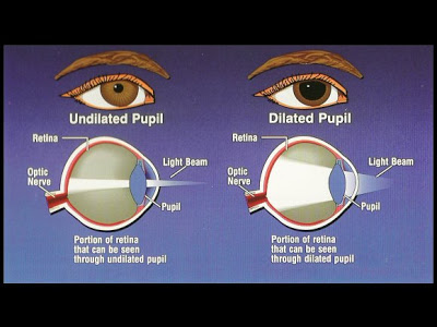
This unit was about the brain and our senses. We first started off with the 
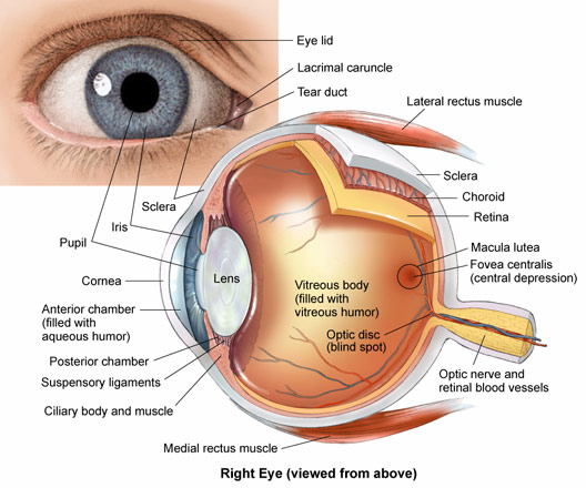
anatomy of the brain and brief overview of what role each part of the brain had. Then, we learned more specifically about the two hemispheres and lobes. The left hemisphere is more detail and fact oriented while the right looks at overall context more. The brain is very malleable and has the ability to reorganize itself to heal from damage or in response to new experiences. Next we touched on all our senses--sight, hearing, touch, taste, and smell--and the specific receptors that are used to sense stimuli. For example, we have thermoreceptors to sense heat and propioreceptors that sense pain. In order for receptors to send signals to the brain, there need to be neurons, which we learned about next. The neural system is organized into the PNS and CNS, each playing an important role in interpreting messages and sending out tasks for motor neurons.
http://climatereview.net/ChewTheFat/wp-content/uploads/2011/11/CNS-vs-PNS.jpg


Readings in this Unit:
"How to become a Superager" by Lisa Barret
This article was about how some older people are able to maintain their brain capability to equal those of younger people in their 20s. She calls those people "superagers". Physically, regular agers' brains degrade after a while of dissuse, The only way to keep the brain at it's peak is to engage it in solving challenges or performing difficult tasks. In our brain lecture we talked about how neurons that fire together, wire together, but here we learn that the opposite is also true. If neurons are not fired, they become disconnected. 

"FIt Body, Fit Brain, and Other Fitness Trends" by Gretchen Reynolds
Reynolds talks about how we can keep our brains in tip-top shape and also explains why each method is necessary. Exercise is linked to brain fitness: improvements in thinking and an increase in the number of neurons. Weight training is particularly important because it leads to fewer lesions in the brain's white matter, which is needed to pass messages to varying parts of the brain. Interestingly enough, in a study about twins, the twin with larger calves had a healthier brain that the twin with punier calves, directly supporting how important weight training is for the brain. Based on lectures, we know why white matter is essential to brain function ( it is full of neurons). If physical and mental exercise helps us in keeping our white matter and increasing the number of neurons in the brain, clearly exercise is something every person should be motivated to do.

"How We Get Addicted" by Michael D. Lemonick
Addictions are defined as "repetitive behaviors in the face of negative consequences", the desire to do something you know is bad for you. Scientists are now able to develop a better understanding of how addictions affect the brain. They found out that drugs stimulate the same brain functions that allowed our ancestors to survive. Exposing ourselves to drugs creates a salience overdrive that creates uncontrollable craving. The reason we are not ALL addicts is because we have a reasoning part of our brain that can tell us that the consequences of addiction are not worth it. The reasoning part of the brain is mostly the prefrontal cortex. As we learned early on in this unit, the brain has many different parts all in charge of different functions. The cortex is involved in making judgements and plans and can override cravings.


Senioritis is hitting me 100% full force and my academic aggression for this class is faltering. I don't do anything unless I have to anymore: I don't cut and paste my notes in until it's due, I take many "breaks" from work. As far as strengths, at least I'm maintaining decent grades and still doing the work for my classes. I also haven't skipped any school even though I have been tempted, so I'll consider that an achievement.  
Based on the previous paragraph, I think it is clear that my goal of staying motivated is on it's way to failure. However, I am maintaining a healthier lifestyle these days. I go running with my friends on a weekly basis and I watch what I eat (can't do that overcarbsumption stuff!). I get adequate sleep, although whether it's because I can't physically stay up anymore like I used to or if it's because I actually want the health benefits of enough sleep is unclear. Go me.

Reflex Lab

In this lab, we conducted multiple experiments to test our reflexes and reactions times. A reflex is an automatic response to a stimulus--often the reaction happens with out a signal being processed by the brain. Instead, the signal is passed to the spinal cord for a faster response to the stimuli. In the first experiment, we tested the reaction of the iris to light. One partner covered her eyes for 2 minutes and then the other partner shined light onto the eye after the 2 minutes. We observed that the pupil constricted in the bright light and dilated when it was dark. The eye adjusts the amount of light that enters the eye to enable us to have optimal vision in all conditions.

https://blogger.googleusercontent.com/img/b/R29vZ2xl/AVvXsEhiGLIwWLJ9FLIUGycrcaoCnECWqH5MwCtGbNDk6gWWbeZ6lcTtutRH_Cvv57lgEVATX_uuzYQdO5GErZXS227zsoa6Tb1IyzemU9qgMxGdXxM6cS_4a1Ri8CUHcUa2S9VTJpZ4GwCtB8o/s400/PupilDilation-grphc.jpg

In the second experiment, we tested the knee jerk reflex by hitting the knee with a knee hammer and observing whether the leg shot up or not in response. Then we performed the same procedure after doing 30 squats to see if the reflex was heightened or slowed. We saw that the reflex was faster and more prominent after the squats, maybe because the blood was pumping and the neurons were more sensitive after exercise.
https://media1.britannica.com/eb-media/62/141362-004-BB0AD15A.jpg

In the third experiment, we tested the blink reflex. A clear sheet was help in from of our faces and a cotton ball was thrown at the face. The person behind the plastic blinked without fail each time the cotton ball was thrown. The body does this because the eyes are a very important part of the body and should be protected from incoming objects to prevent damage.
For the fourth experiment, the plantar reflex was tested. A pen was dragged along the sole of someone's foot. In response to the stimulus, the toes scrunched up. This happened for both partners. If the nervous system is damaged, the toes would have spread apart and up. Perhaps this reflex is to protect the foot from too much damage if you step on something by lifting most of the foot away from the ground.
In the fifth and final experiment, we tested our reaction time for when we are focused vs. unfocused. One partner held a meter stick and the other put her had near the bottom of the ruler. The first partner drops the ruler and the second is supposed to catch it. The inches in which partner 2 catches the meter stick was recorded and translated into a time measurement to approximate how fast the reaction time was. For the unfocused version, the same experiment was performed except the person catching the ruler was not texting at the same time. My average reaction time when focused was 0.11 seconds while my unfocused time was 0.193. Caroline's times had a similar pattern, her first average being 0.13 and the second being 0.2 seconds. The distracted response time was slower because the brain is now multitasking and had to split the focus between two things. 


H2OnTheGo (post2)


image1.JPGSo far, I've drawn up concept designs and did a little research about
    how the electronic parts of my bottle
will work.


The bottle will be a sippy cup type mechanism for convenience. Currently, tech savvy bottle all have a screw off cap, but based on some friends I questioned and what I've observed, people prefer not to have to expend the energy to actually screw off and on caps.As shown flip off part of the cap will show things like the current temperature and time.An extra possibility is to add a detachable infuser because people will more willingly drink flavored water and the plastic it would take to make the infuser would cost very little. My infuser design is also unique and new in that it is deconstructable for easy cleaning.
The bracelet will be a silicon band that gradually flattens at the ends to where the disc magnets will be placed to make the band adjustable-fits all sizes. A button off to the side of the body will enable the user to turn on and off the auditory reminder to drink water. The four water droplets will glow through the silicon covering. Based on the temperature sensor in the bottle cap, the number of glowing droplets will change--one being drink an average amount of water, four being water intake should be increased by a lot (maybe 2X or something like that).

image2.JPGThere will be a silicon strip in the middle part of the glass bottle for grip.


image3.JPG


I haven't really had any setbacks so far, Next, I plan to research the costs of materials.

Thursday, March 9, 2017

Brain Dissection

In this lab, we made cuts into the brain so that we could observe the many parts of the brain by looking at cross sections. We saw how the brain was physically divided into the left and right hemispheres and identified various other parts like the cerebellum and thalamus. In the cross section of the cerebrum, we were able to differentiate white matter from gray matter. The brain shares some similarities to the heart in that his had a protective layer around it that held the brain together, called the meninges.

Question 1: Take a picture with your pins in place. Draw a detailed sketch of the brain and label each of the structures.

drawing of the whole brain from the left side
whole brain viewed from left side

Question 2: What is the function of each of these structures?
  • Cerebrum-integrates the messages received in the brain
  • Cerebellum-motor control
  • brain stem-controls basic functions like breathing, digestion; filters sensory information
Question 3: What is the function of myelin in a neuron?
The myelin insulates and increases the speed of nerve impulses in the neuron.

Question 4: Picture and drawing of cross section with labels.
drawing of cross section along the medial plane

cross section of brain along medial plane

Question 5: What is the function of each of the structures you pinned in step 8?
  • thalamus- sorts data, autonomic activities, maintains consciousness
  • optic nerve- connects eye to brain for sight
  • medulla oblongata- controls heartbeat, breathing
  • pons- contains bundles of axons that connect parts of the brain with one another
  • midbrain- connects nerve impulses from cerebrum to spinal cord, medulla, and pons
  • corpus callosum-connects right and left hemispheres of the brain with nerves
  • hypothalamus- maintains homeostasis
Question 6: Make a detailed drawing of your cross section and take a pic.
drawing of ca cross section of the cerebrum
cross section of cerebrum


Monday, March 6, 2017

Sheep Eye Dissection Analysis

http://www.biographixmedia.com/human/eye-anatomy.jpg
On the exterior of the eye, there are rectus muscles that helps the eye turn. The white of the eye, which is actually really tough, is the sclera. 
When a person sees, light first passes through a hard shell called the cornea. It helps focus light and protects the eye against the dust and other particles. The iris, which is behind the cornea, controls the amount of light that enters through the pupil and hits the lens. Between the lens and the cornea is the aqueous humor, a liquid that maintains the pressure of the eyeball. The light is bent by the lens to focus the light onto the retina at the back of the eye. Ciliary muscles alters the curve of the lens to help focus the light. In the retina, rods and cones detect color and gray scale to send messages to the brain. Where the optic nerve starts, we have a blind spot where there is a lack of rods and cones. Under the retina is the choroid that supplies the eye with blood. The optic nerves and blood vessels funnel together at the back of the eye. Visual messages are then sent to the brain. Vitreous humor in the main cavity of the eye ball maintains the shape of the eye.